
Goat Anti-HIF1α Polyclonal IgG Antibody: A Key Tool for Hypoxia Research
In biomedical science, detecting molecular signals that regulate oxygen homeostasis is essential for understanding diseases like cancer, ischemia, and chronic inflammation. One of the most widely used reagents for this purpose is the Goat Anti-HIF1α Polyclonal IgG Antibody, which enables researchers to detect Hypoxia-Inducible Factor 1-alpha (HIF-1α)—a protein that plays a crucial role in how cells respond to low oxygen.
This article explores what the antibody is, how it works, and how it supports cutting-edge research in immunology, oncology, and cell biology.
What Is HIF-1α?
HIF-1α (Hypoxia-Inducible Factor 1-alpha) is a transcription factor that regulates gene expression in response to hypoxia, or low oxygen conditions. It activates genes involved in metabolism, angiogenesis, cell survival, and adaptation to oxygen deficiency. HIF-1α is central to studies on tumor progression, stroke, wound healing, and cardiovascular disorders.
You can learn more about HIF-1α through the National Library of Medicine (NCBI Gene), and its regulatory functions are well-documented by the National Cancer Institute.
Why Use a Goat Anti-HIF1α Polyclonal IgG Antibody?
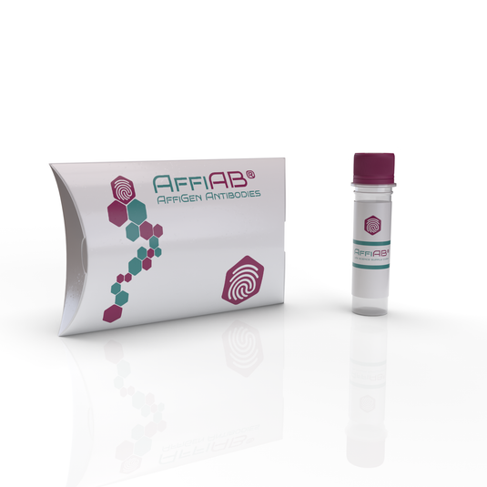
The Goat Anti-HIF1α Polyclonal IgG Antibody is produced by immunizing goats with HIF-1α protein fragments. This leads to the generation of polyclonal antibodies—a diverse pool that binds to multiple epitopes of the HIF-1α protein. This diversity makes the antibody highly sensitive and ideal for various detection methods.
Key benefits of using a polyclonal goat antibody include:
-
High binding affinity to target proteins
-
Better detection of conformationally variable epitopes
-
Enhanced signal intensity in Western blot, ELISA, IHC, and IF applications
A great resource on polyclonal antibodies is available from Harvard University’s Core Facilities and a comparison guide from University of Iowa Central Microscopy.
Applications in Research
The Goat Anti-HIF1α antibody is essential in many labs studying cellular hypoxia, as well as:
1. Cancer Biology
Research at MD Anderson Cancer Center and University of Michigan Rogel Cancer Center demonstrates how HIF-1α promotes tumor angiogenesis and metastasis under low oxygen.
2. Stroke and Ischemia Studies
Groups like the National Institute of Neurological Disorders and Stroke (NINDS) use antibodies targeting HIF-1α to monitor neural hypoxia and damage during cerebral ischemia.
3. Wound Healing and Inflammation
At Stanford University, researchers track HIF-1α to investigate how wounds heal in oxygen-deficient tissue.
4. Metabolic Disorders
NIH-funded studies have linked HIF-1α regulation to glucose metabolism and insulin resistance, highlighting its role in diabetes research.
Techniques and Protocols
The antibody is commonly used in techniques such as:
-
Western blotting – For detecting HIF-1α protein in lysates
-
Immunohistochemistry (IHC) – For localizing HIF-1α in tissue sections
-
Immunofluorescence (IF) – For visualizing HIF-1α in cells
-
ELISA – For quantifying HIF-1α levels in biofluids
Protocols for these applications are published by institutions like University of California, Davis and Johns Hopkins Pathology Core.
The Centers for Disease Control and Prevention (CDC) also provides general guidance for immunoassays and lab safety.
Antibody Validation and Specificity
Because HIF-1α is often present at low levels under normoxia, high specificity is vital. Validation is typically done through knockdown or overexpression models, which you can read about at NCBI’s Antibody Validation Portal and NIH’s Office of Research Infrastructure Programs (ORIP).
University of California, San Francisco (UCSF) and University of Chicago labs use such models to verify antibody reliability in both human and mouse studies.
Structure and Format
This antibody is typically supplied as:
-
Whole IgG purified by Protein G affinity chromatography
-
Host species: Goat
-
Target species: Human (cross-reactivity with mouse and rat possible)
-
Storage: Long-term at -20°C, avoid repeated freeze-thaw cycles
You can read about proper antibody storage in resources from University of Minnesota and Purdue University research cores.
ELISA Optimization with Goat Anti-HIF1α IgG
When using the antibody in HIF1α ELISA, it is important to:
-
Use standard curves for quantification
-
Include positive and negative controls
-
Optimize incubation times and blocking buffers
University of Florida Biotech Core and Cornell Veterinary Core offer step-by-step ELISA optimization guides.
Real-World Examples
-
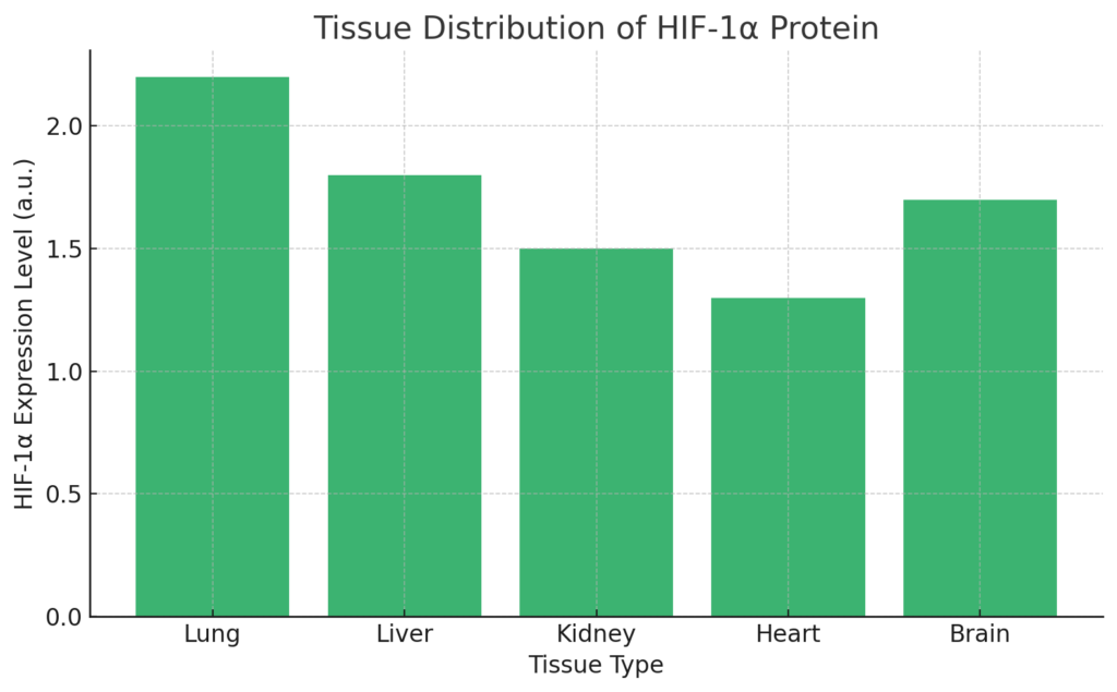
Environmental Health Studies: EPA-funded studies assess how pollutants influence HIF-1α expression in lung tissues.
-
Developmental Biology: UC Berkeley labs use this antibody to explore oxygen-regulated gene expression in embryos.
-
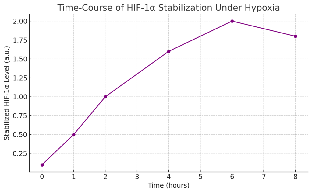
High-Altitude Research: University of Colorado Boulder tracks HIF-1α in studies of altitude adaptation and blood oxygenation.
Public Resources and Education
If you’re new to the field or want to understand the basics of antibody-based research:
For protocol repositories:
Conclusion
The Goat Anti-HIF1α Polyclonal IgG Antibody is a trusted tool for detecting one of the most important regulators of the hypoxic response in human biology. From cancer to inflammation and wound repair, this antibody supports discoveries that shape how scientists view oxygen’s role in health and disease.
With wide use across academic institutions and support from federal research agencies like NIH, CDC, and EPA, this antibody continues to drive progress in both basic science and applied research.

History of ARKdb
Using ARKdb
Matrix 3D Cell Culture Gel – Rigid: A High-Stiffness Substrate for Advanced Biomedical Research
About Author
