
Optimizing Giemsa Stain PR80 for Accurate Cytogenetic Analysis in Cancer Diagnostics
Cytogenetic analysis plays a pivotal role in cancer diagnostics, as it provides essential insights into chromosomal abnormalities linked to the onset and progression of cancer. Giemsa staining, one of the most common and effective techniques for chromosome banding, has been in use for decades to visualize chromosomal structures. The PR80 variant of Giemsa stain, developed for high-resolution chromosomal analysis, has become an indispensable tool in modern cancer diagnostic laboratories. This article explores the optimization of Giemsa Stain PR80 to improve cytogenetic analysis accuracy, with a focus on cancer detection.
Introduction to Giemsa Staining and Its Role in Cytogenetics
Cytogenetics is the study of chromosomes, particularly the structure, number, and behavior of chromosomes during cell division. It provides crucial information about genetic diseases, birth defects, and cancer (1). Giemsa staining is one of the most reliable methods used to examine chromosomes. By staining chromosomes in a way that differentiates regions with varying DNA content, this technique allows cytogeneticists to visualize individual chromosomal bands that are otherwise invisible under a microscope (2).
Giemsa Stain PR80, a more refined version of the conventional Giemsa stain, is known for providing clearer and sharper banding patterns, which are essential for identifying minor chromosomal alterations. These alterations can have significant implications in the diagnosis and treatment of cancer, making the optimization of Giemsa Stain PR80 an essential step in improving the sensitivity and accuracy of cytogenetic analysis (3).
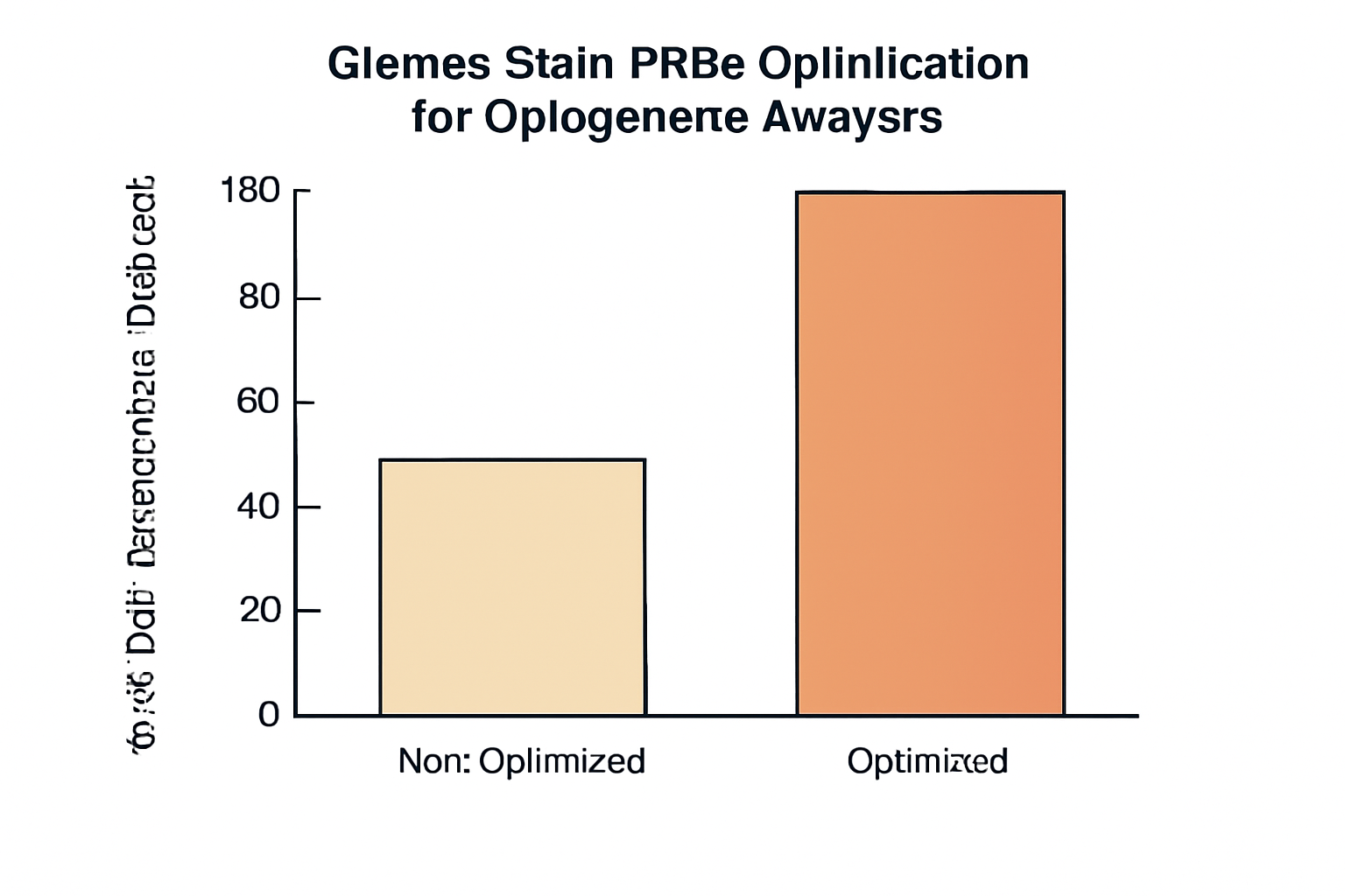
The Importance of Optimizing Giemsa Stain PR80 in Cancer Diagnostics
In cancer diagnostics, accurate cytogenetic analysis is crucial for detecting chromosomal abnormalities that are often associated with various forms of cancer. These abnormalities may include translocations, deletions, duplications, and other structural changes that can lead to malignancies (4). Giemsa Staining, by enabling the identification of these changes, provides invaluable information that assists in determining the prognosis and appropriate treatment for cancer patients.
However, the sensitivity of cytogenetic analysis can be compromised if the staining process is not optimized. For example, insufficient stain concentration, improper staining duration, or unsuitable washing procedures can result in unclear banding patterns that fail to highlight chromosomal anomalies (5). This issue is especially critical in cancer diagnostics, where small chromosomal changes often have major implications for diagnosis and treatment (6).
Key Factors for Optimizing Giemsa Stain PR80
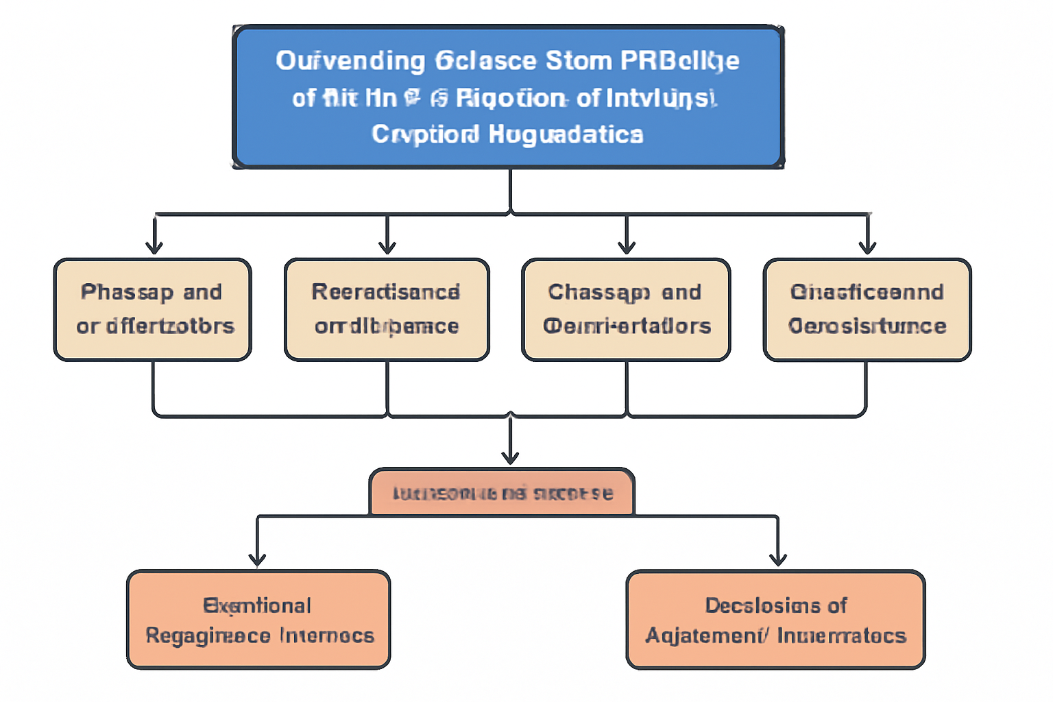
To achieve accurate and high-quality cytogenetic analysis, optimizing Giemsa Stain PR80 involves fine-tuning several key factors in the staining procedure. The main factors influencing the quality of Giemsa staining include the sample preparation process, stain concentration, staining time, temperature, and post-staining washing and dehydration steps.
1. Sample Preparation
The preparation of the sample is the first and most important step in the staining process. Chromosomal analysis requires that cells be in metaphase, as chromosomes are most compact and easily visible during this stage (7). Sample preparation also involves ensuring that the cells are properly fixed and mounted on glass slides. If cells are not adequately fixed, they may shrink or distort, leading to blurry or fragmented chromosome images that make it difficult to detect subtle chromosomal abnormalities (8).
The quality of cell culture, fixation methods, and slide preparation all play a significant role in the final outcome of the cytogenetic analysis. Proper fixation ensures that the chromosomes maintain their integrity and that the staining process produces clear results (9). Additionally, keeping the slides clean and free from contamination is essential to avoid interference in the visualization process.
2. Stain Concentration
The concentration of the Giemsa stain is another crucial factor in obtaining optimal results. If the concentration of the stain is too low, the chromosomes will not absorb enough stain, leading to faint and unclear banding patterns. On the other hand, a high concentration of Giemsa stain may lead to excessive background staining, which can obscure the chromosomal bands (10).
For Giemsa Stain PR80, the ideal concentration is typically around 5%, although this may vary depending on the specific requirements of the sample and the laboratory protocols (11). Adjusting the concentration based on these parameters is critical for obtaining sharp and well-defined banding patterns.

3. Staining Time and Temperature
Staining time and temperature are also key factors that influence the final quality of the Giemsa staining. If the staining is too brief, the chromosomes will not be stained adequately, leading to poor visibility. Alternatively, over-staining can result in excessive background color that interferes with the detection of chromosomal bands (12).
For Giemsa Stain PR80, the recommended staining time is typically between 15 to 20 minutes at room temperature. However, certain protocols may require minor adjustments to achieve the desired level of staining intensity. Temperature also plays a role, as higher temperatures tend to speed up the staining process, while lower temperatures may slow it down. It is essential to monitor the temperature carefully, as extreme fluctuations can affect the quality of the staining (13).
4. Post-Staining Washing and Dehydration
After staining, it is necessary to thoroughly wash the slides to remove any excess stain. Inadequate washing can result in excessive background staining, which diminishes the contrast between the chromosomes and the background (14). This is especially important in cancer diagnostics, where clarity and precision are required to detect subtle chromosomal changes.
The post-staining dehydration process is equally important. Dehydration helps preserve the chromosomal structure and ensures that the stained chromosomes are visible under the microscope. A graded alcohol series is typically used for this purpose, and it is essential to ensure that the slides are fully dried before proceeding to imaging (15).
Applications of Optimized Giemsa Staining in Cancer Diagnostics
Optimizing Giemsa Stain PR80 is critical for detecting chromosomal abnormalities associated with various types of cancer. Many cancers, including leukemia, lymphoma, and breast cancer, are characterized by specific chromosomal translocations or mutations that can significantly influence the course of the disease (16). By optimizing the Giemsa staining process, it becomes easier to identify these chromosomal abnormalities, facilitating early diagnosis and personalized treatment strategies.
In particular, Giemsa Staining is vital for detecting structural changes in the chromosomes, such as the Philadelphia chromosome in chronic myelogenous leukemia (CML). This chromosomal translocation is a hallmark of CML and can be detected with high sensitivity when the Giemsa staining protocol is properly optimized (17).
Moreover, in solid tumors such as breast cancer, ovarian cancer, and lung cancer, the use of Giemsa Staining PR80 allows for the detection of gene amplifications, deletions, and other chromosomal changes that influence prognosis and treatment decisions (18).
Enhancing Sensitivity and Resolution with Giemsa Stain PR80
One of the primary advantages of Giemsa Stain PR80 is its ability to detect even minor chromosomal abnormalities that may be missed using conventional staining methods. To further enhance sensitivity, cytogeneticists can combine Giemsa staining with other techniques, such as fluorescence in situ hybridization (FISH), which allows for the identification of specific gene regions or mutations (19).
FISH can be particularly useful when analyzing chromosomal regions that are difficult to distinguish using Giemsa staining alone. By combining these two techniques, it becomes possible to conduct a more comprehensive cytogenetic analysis, leading to more accurate detection of chromosomal abnormalities (20).
Conclusion
Optimizing Giemsa Stain PR80 is essential for achieving accurate cytogenetic analysis, particularly in cancer diagnostics. By adjusting key parameters such as stain concentration, staining time, and sample preparation, cytogeneticists can improve the resolution and clarity of the chromosomal bands, making it easier to detect chromosomal abnormalities. This enhanced detection is vital for the early diagnosis and treatment of various cancers.
With the increasing reliance on genetic and chromosomal analysis in oncology, the role of optimized Giemsa Staining remains central to advancing cancer diagnosis and improving patient outcomes. For further information on cytogenetic techniques and their role in cancer research, refer to resources such as the National Institutes of Health (NIH), National Cancer Institute (NCI), and Centers for Disease Control and Prevention (CDC).
References:

Aspergillus fumigatus IgG ELISA Kit: An Essential Tool for Detecting Fungal Infections
Goat Anti-mNectarine Polyclonal IgG Antibody: Applications and Research Insights
History of ARKdb
About Author
