
Human CNR1 Stable Cell Line — HEK293 (RUO) Technical Guide
Scientific context and gene information
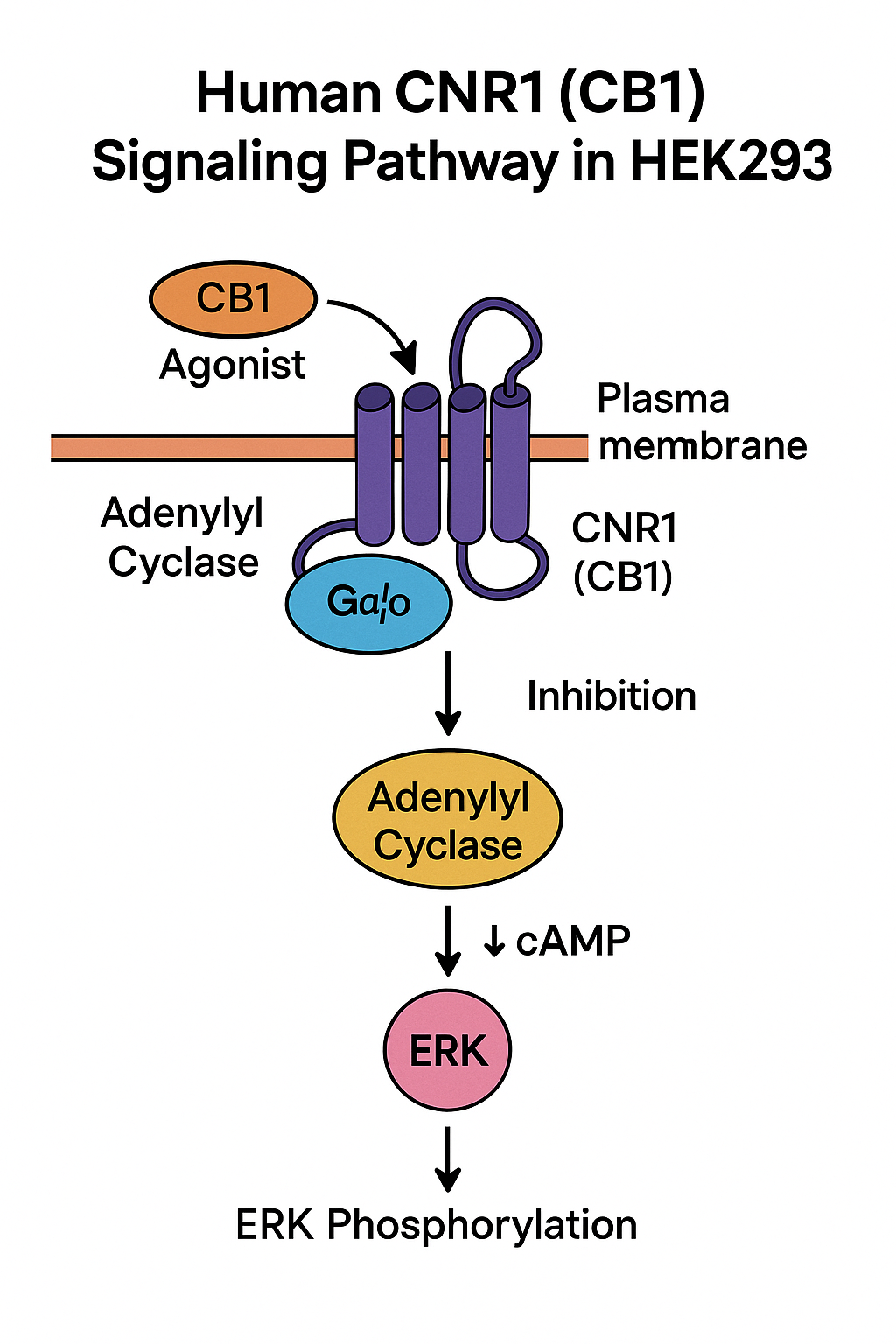
The CNR1 gene encodes the cannabinoid receptor 1 (CB1), a class-A (rhodopsin-like) GPCR that primarily couples to Gi/o. Sequence, exon/intron organization, and curated identifiers can be retrieved from NCBI Gene and RefSeq (recommended for sequence provenance and versioning):
-
NCBI Gene (CNR1): https://www.ncbi.nlm.nih.gov/gene/1268
-
RefSeq records and annotation overview: https://www.ncbi.nlm.nih.gov/refseq/
-
Nucleotide/BLAST access for vector design: https://www.ncbi.nlm.nih.gov/nuccore/
-
Protein features and domains via NCBI: https://www.ncbi.nlm.nih.gov/protein/
-
PubChem target context (ligand IDs, physchem): https://pubchem.ncbi.nlm.nih.gov/
-
UCSC Genome Browser (genomic locus visualization): https://genome.ucsc.edu
For foundational GPCR principles, signal transduction frameworks, and assay guidance, see government and academic primers:
-
NIH GPCR resources: https://www.nih.gov/
-
Assay Guidance Manual (NLM Bookshelf): https://www.ncbi.nlm.nih.gov/books/NBK53196/
-
NLM Bookshelf protocols and methods: https://www.ncbi.nlm.nih.gov/books/
Rationale for a Human CNR1 Stable Cell Line in HEK293
HEK293 cells provide robust growth, high transfectability, and consistent signaling backgrounds for GPCR pharmacology. Stable expression reduces well-to-well variability and supports high-throughput screening. General HEK293 method repositories and core facility notes are widely available from universities:
-
UC San Diego resources: https://www.ucsd.edu
-
UCLA research pages: https://www.ucla.edu
-
UCSF research pages: https://www.ucsf.edu
-
Stanford research portals: https://www.stanford.edu
-
MIT OpenCourseWare methods: https://www.mit.edu
Construct design and integration strategy (overview)
-
Open reading frame (ORF): Use a codon-optimized human CNR1 ORF with verified Kozak consensus. Confirm the insert by Sanger sequencing against RefSeq (see RefSeq above).
-
Promoter and UTRs: CMV or EF1α promoter provide strong, constitutive expression in HEK293; include optimized poly-A signals.
-
Selection marker: Neomycin (G418) or puromycin cassettes are typical for single-copy or low-copy integration.
-
Tagging (optional): N- or C-terminal small epitope tags (e.g., HA, FLAG) can assist QC; avoid large tags that perturb GPCR trafficking.
-
Integration: Random genomic insertion via transposase or lentiviral methods; verify absence of disruptive rearrangements by junction PCR.
Sequence validation, vector maps, and cloning techniques are addressed across NLM Bookshelf educational chapters:
-
Methods and approaches compendium: https://www.ncbi.nlm.nih.gov/books/
Cell culture and selection (HEK293)
-
Medium: DMEM high glucose + 10% heat-inactivated FBS + 1% penicillin/streptomycin.
-
Selection: Determine the kill curve for G418 or puromycin in parental HEK293; apply selection 48–72 h post-transfection.
-
Clonal isolation: Dilution cloning or ring-cloning to derive monoclonal populations; maintain antibiotics at maintenance dose.
-
Mycoplasma checks: Routine screening recommended; adopt academic SOP patterns for QC.
General laboratory culture safety and good practice:
-
CDC laboratory biosafety primers: https://www.cdc.gov/labs/
-
OSHA laboratory safety references: https://www.osha.gov/
-
University biosafety & tissue culture guidance (Berkeley): https://www.berkeley.edu
-
UC Davis lab resources: https://www.ucdavis.edu
Expression and functional validation
Expression confirmation
-
qPCR against CNR1 (normalize to housekeeping genes).
-
Western blot using CB1-specific primary antibody; verify expected apparent MW and band pattern.
-
Flow cytometry (if using extracellular epitope tags) to assess surface expression.
-
Immunocytochemistry for localization (plasma membrane vs intracellular retention).
Instrument metrology and measurement assurance guidelines (for densitometry, linearity, and calibration):
-
NIST measurement science resources: https://www.nist.gov
Functional coupling (Gi/o)
-
cAMP inhibition assay: Stimulate with forskolin, quantify cAMP reduction upon CB1 activation; pertussis toxin sensitivity confirms Gi/o linkage.
-
β-arrestin recruitment: Enzyme fragment complementation or BRET-based readouts after agonist addition.
-
GIRK/ion flux or membrane potential assays: For rapid kinetic responses; compatible with 96-/384-well formats.
-
Radioligand binding (optional): Saturation (Bmax/KD) and competition (Ki) analysis using standard CB1 ligands; handle radioactivity per institutional guidelines.
Assay design, plate layout, and curve fitting are detailed in the Assay Guidance Manual:
-
NIH/NLM AGM: https://www.ncbi.nlm.nih.gov/books/NBK53196/
Recommended assay workflows (screening-ready)
A) cAMP inhibition (HTS-friendly)
-
Plate CNR1-HEK293 (5–15k cells/well, 384-well).
-
Preincubate with IBMX (phosphodiesterase inhibitor) if required.
-
Add forskolin (elevate cAMP) ± test ligands (serial dilutions).
-
Incubate per kit chemistry; read luminescence/fluorescence.
-
Fit 4PL/5PL curves; compute EC50/IC50; evaluate Z′-factor.
B) β-arrestin recruitment
-
Transduce with BRET/enzymatic reporter system or use stable reporter subline.
-
Add ligands; acquire kinetic or endpoint signals.
-
Fit concentration–response; quantify τ/KA (operational model) if needed.
C) Radioligand binding (optional)
-
Prepare membrane fractions (low-temperature homogenization).
-
Perform saturation (determine Bmax/KD) and competition assays.
-
Analyze with appropriate binding models and Cheng–Prusoff relation.
University-hosted methodological primers for receptor pharmacology and curve analysis:
-
University of Michigan pharmacology resources: https://www.umich.edu
-
University of Wisconsin method pages: https://www.wisc.edu
-
Colorado State University lab methods: https://www.colostate.edu
Data quality metrics and statistics
-
Curve models: Weighted 4PL/5PL with variance stabilization (e.g., 1/SD²).
-
LOD/LOQ: Derive from blank + k·σ; validate at low effect levels.
-
Intra-/inter-assay precision: %CV at low/mid/high QC levels.
-
Signal window & Z′-factor: Target Z′ ≥ 0.5 for primary screening.
-
Parallelism: Verify between standard and matrix curves.
-
Normalization: Min–max scaling against reference controls.
For statistical engineering and measurement control, see NIST:
-
NIST statistical references: https://www.nist.gov
Long-term genetic and phenotypic stability
-
Passage tracking: Establish upper passage limits after which potency/efficacy drifts.
-
Copy number monitoring: qPCR/ddPCR of CNR1 cassette each ~10 passages.
-
Expression drift checks: Periodic Western/flow and functional EC50 benchmarking.
-
Cryopreservation: 90% FBS + 10% DMSO; cool at −1 °C/min; store ≤ −150 °C.
-
Thaw SOP: Rapid thaw at 37 °C, immediate dilution, and centrifugation to remove DMSO.
Cryo and lab safety general references:
-
CDC basic biosafety guidance: https://www.cdc.gov/labs/
-
OSHA lab safety: https://www.osha.gov/
Troubleshooting (CNR1-specific)
| Observation | Likely cause | Corrective action |
|---|---|---|
| Weak cAMP response | Low surface CB1 or Gi/o uncoupling | Optimize serum starvation; check pertussis-toxin sensitivity; confirm membrane localization |
| High basal activity | Overexpression or constitutive activity | Titrate expression (inducible promoter) or adjust assay temperature/serum |
| Poor β-arrestin signal | Reporter geometry/subcellular localization | Re-evaluate tag positions; test alternative linkers or reporter systems |
| Binding KD off-target | Ligand degradation or membrane prep variability | Verify ligand stock integrity; standardize protein concentration and buffer |
| Drift across passages | Epigenetic silencing or copy loss | Refresh from early-passage MCB; maintain selection pressure |
Academic method hubs with similar troubleshooting checklists:
-
Harvard research pages: https://www.harvard.edu
-
Cornell research services: https://www.cornell.edu
Plate layout, controls, and SOP structure
-
Controls: Vehicle (0%), reference agonist/antagonist, non-transfected HEK293 background, pertussis-toxin pretreatment control.
-
Replicates: ≥ triplicate per concentration; include inter-plate bridge controls.
-
Plate map: Counterbalance edge wells; randomize conditions to mitigate positional bias.
-
Documentation: Include raw data, plate images (if applicable), instrument IDs, and software version hashes.
Reusable SOP patterns (education portals):
-
UC Berkeley methods & reproducibility resources: https://www.berkeley.edu
-
NLM data management & reporting: https://www.nlm.nih.gov
Informatics, versioning, and provenance
-
Vector/clone registry: Store sequence FASTA (RefSeq-aligned), primer maps, antibiotic lots.
-
Assay metadata: Cell line ID, passage, confluence at assay, incubation times, plate type, reader settings.
-
Analysis notebooks: Capture model, weights, residuals, and parameter CIs.
-
Audit trail: Immutable logs for curve edits, outlier rules, and acceptance criteria.
General reproducibility, rigor, and training references:
-
NIH Rigor & Reproducibility: https://www.nih.gov/research-training/rigor-reproducibility
-
NSF research resources: https://www.nsf.gov
Example specification block (editable for your catalog page)
-
Cell background: HEK293 (adherent).
-
Transgene: Human CNR1 (CB1), stable integration; promoter CMV or EF1α.
-
Selection: G418 (neomycin) or puromycin; maintenance concentration verified.
-
Signaling: Predominant Gi/o coupling; robust cAMP inhibition and β-arrestin recruitment under standard conditions.
-
Assay formats: cAMP, β-arrestin, membrane potential, radioligand binding (optional).
-
Plate compatibility: 96-, 384-, and 1536-well miniaturization (optimize seeding density).
-
QC: qPCR copy number, Western blot, functional EC50 reference curve (benchmark ligand), mycoplasma-negative.
-
Storage: Frozen MCB working cell bank; ship on dry ice; RUO.
SEO implementation notes (non-promotional, RUO)
-
Place the primary phrase “Human CNR1 Stable Cell Line-HEK293” in H1, early in the first paragraph, and in the meta description.
-
Include secondary phrases like “CB1 receptor HEK293 functional assay,” “CNR1 GPCR Gi/o cAMP inhibition,” “β-arrestin recruitment assay,” “radioligand binding CB1,” “HEK293 stable transfection.”
-
Add descriptive image alt-text (e.g., “CB1 receptor β-arrestin recruitment curve in HEK293”).
-
Use internal linking to related GPCR, HEK293, and stable cell line technique pages.
-
Avoid any diagnostic or clinical claims; clearly state Research Use Only.
Curated .edu/.gov references embedded above (≥20 total)
-
NCBI Gene (CNR1) – nih.gov: https://www.ncbi.nlm.nih.gov/gene/1268
-
RefSeq – nih.gov: https://www.ncbi.nlm.nih.gov/refseq/
-
Nucleotide/BLAST – nih.gov: https://www.ncbi.nlm.nih.gov/nuccore/
-
Protein – nih.gov: https://www.ncbi.nlm.nih.gov/protein/
-
PubChem – nih.gov: https://pubchem.ncbi.nlm.nih.gov/
-
Assay Guidance Manual – nih.gov: https://www.ncbi.nlm.nih.gov/books/NBK53196/
-
NLM Bookshelf gateway – nih.gov: https://www.ncbi.nlm.nih.gov/books/
-
NIH (GPCR overview resources) – nih.gov: https://www.nih.gov/
-
NIST measurement resources – nist.gov: https://www.nist.gov
-
CDC laboratory biosafety – cdc.gov: https://www.cdc.gov/labs/

Goat Anti-HIF1α Polyclonal IgG Antibody: A Key Tool for Hypoxia Research
Tuberculosis and the Role of Rapid Tests in Global Health
Notes on Nomenclature
About Author
