
Programmed Cell Death Protein 4 (PDCD4) ELISA — Technical Guide for Research Laboratories
Molecular background (for assay designers)
Programmed cell death protein 4 (PDCD4) is a translation regulator encoded by PDCD4 and conserved across vertebrates. For sequence, exon structure, and known isoforms, consult NCBI Gene and RefSeq entries (e.g., PDCD4, Homo sapiens) on NCBI and NLM resources:
• NCBI Gene overview: ncbi.nlm.nih.gov/gene
• RefSeq and GenPept records: ncbi.nlm.nih.gov/refseq
• Conserved Domains / CDD for MA-3 domains: ncbi.nlm.nih.gov/Structure/cdd
• FASTA and CDS retrieval via Nucleotide: ncbi.nlm.nih.gov/nuccore
• UCSC Genome Browser gene context: genome.ucsc.edu
Protein-level characteristics that influence ELISA development include epitope accessibility, post-translational modifications (PTMs), and potential proteolysis. For curated protein features and literature mapping, see NCBI Bookshelf protocols and handbooks that outline antibody selection and antigen design strategies:
• NLM Bookshelf (assay development chapters): ncbi.nlm.nih.gov/books
Assay principle: PDCD4 sandwich ELISA (RUO)
A typical sandwich ELISA for PDCD4 uses a capture antibody immobilized on a high-binding polystyrene 96-well microplate and a biotinylated or enzyme-conjugated detection antibody recognizing a non-overlapping epitope. The signal is generated through HRP/TMB or AP/pNPP systems and read at 450 nm (or 405 nm for AP) on a calibrated microplate reader. For foundational ELISA concepts and microplate practice, reference university and government resources:
• UC Davis immunoassay primers: ucdavis.edu
• UWisc laboratory methods pages: wisc.edu
• Colorado State University lab notes (ELISA overview): colostate.edu
• NIST general measurement assurance concepts: nist.gov
• NIH Assay Guidance Manual (AGM): ncbi.nlm.nih.gov/books/NBK53196
Scope: Research Use Only (RUO). Do not interpret results outside controlled laboratory studies.
![AffiELISA® Mouse Programmed cell death protein 4 ELISA [ Pdcd4]](https://affigen.com/cdn/shop/files/5BAFG-E4345_5D_20AffiELISA_C2_AE_20Cattle_20IFNg_20Kit_20High-Resolution_20Interferon_20Gamma_20ELISA_20Detection_f0b190a7-c2bb-4b09-bc2d-e1015cb4a2da_535x.png?v=1712851655)
Reagents, materials, and documentation
-
High-binding microplates (clear, flat-bottom 96-well).
-
Capture antibody (epitope A) and detection antibody (epitope B) validated for non-overlap.
-
Recombinant PDCD4 standard calibrated by protein mass (µg/mL to pg/mL), with verified purity (SDS-PAGE).
-
Blocking buffer (BSA or casein; low IgG background).
-
Assay diluent optimized for matrix components (detergents, proteins, salts).
-
Substrate (TMB/H₂O₂ for HRP) and stop solution (acid).
-
Plate washer or validated manual wash method.
-
Reader with traceable wavelength accuracy and linearity verification (see NIST guidance on instrument metrology: nist.gov).
-
SOPs and batch records aligned with NIH AGM best practices: Assay Guidance Manual.
For general SOP examples and documentation structures, many university pages are useful starting points for internal templates:
• MIT OpenCourseWare lab techniques: mit.edu
• Stanford laboratory safety & methods portals: stanford.edu
• University of Michigan lab method gateways: umich.edu
Sample types, collection, and handling (research matrices)
Common research matrices include clarified cell lysates and tissue homogenates prepared with protease-phosphatase inhibitors. When exploring soluble fractions, define buffer composition, ionic strength, and detergents to maintain epitope integrity. For generic biospecimen handling and biorepository practices, see:
• NCI biospecimen best practices (general): cancer.gov
• CDC general laboratory biosafety references (for safe handling): cdc.gov/labs
Avoid freeze-thaw cycles. Establish stability windows at 2–8 °C and ≤ −70 °C for long-term storage via designed stability studies per AGM guidance.
Plate layout and controls
Design a plate map with: blank, zero, standards (8–10 points, serial 1:2 or 1:3), QC controls (low/mid/high), unknowns, matrix spikes, and dilution linearity checks. Guidance for calibration hierarchy and QC principles can be found via:
• NIST measurement quality resources: nist.gov
• NIH AGM sections on standard curves and QC: ncbi.nlm.nih.gov/books/NBK53196
Recommended procedure (example, sandwich HRP/TMB)
-
Coat: 1–5 µg/mL capture antibody in carbonate buffer, 100 µL/well, 2–16 h at 2–8 °C.
-
Block: 200–300 µL/well blocking buffer, 1 h, RT.
-
Add standards/QCs/samples: 100 µL; incubate 1–2 h, RT or per optimization.
-
Wash: 3–5× with PBS-T.
-
Detect: add detection antibody (0.1–1 µg/mL) or streptavidin-HRP if biotinylated; incubate 30–60 min.
-
Wash: 3–5×.
-
Develop: add TMB (monitor color).
-
Stop: add acid stop; read at 450 nm with 570–620 nm reference if available.
-
Document: retain raw OD, plate map, curve fit, and residual plots per AGM data integrity guidance.
For general ELISA technique primers and troubleshooting checklists, refer to:
• Harvard laboratory method portals: harvard.edu
• UC Berkeley research method pages: berkeley.edu
Calibration models and reportable range
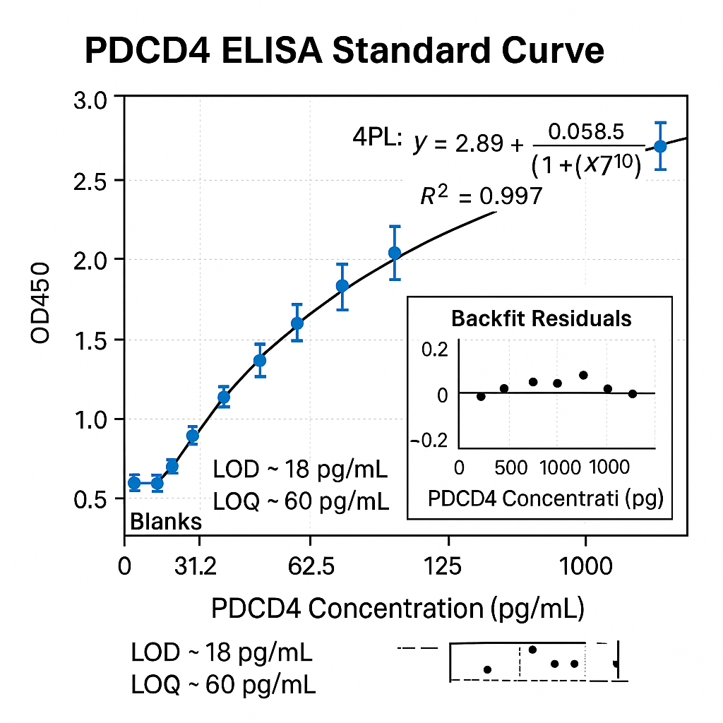
Fit the standard curve using weighted 4PL or 5PL regression with 1/Y² or 1/SD² weighting when heteroscedasticity is present. Determine LOD (blank + 3.3σ) and LOQ (blank + 10σ) using replicate blanks and low-level standards. See rigorous curve-fitting and validation considerations in:
• NIH Assay Guidance Manual (curve fitting, LOD/LOQ): ncbi.nlm.nih.gov/books/NBK53196
• NIST Statistical Engineering Division primers: nist.gov
Validation panels (RUO)
Precision:
-
Intra-assay CV%: evaluate ≥ 20 replicates at low/mid/high.
-
Inter-assay CV%: evaluate across ≥ 3 days, operators, reagent lots.
Accuracy / Trueness (spike-and-recovery):
-
Spike recombinant PDCD4 into matrix at ~80/50/20 % of the mid-standard; acceptable mean recovery typically 80–120 % (define internal limits).
Linearity of dilution / parallelism:
-
Serially dilute matrix samples to assess matrix-specific interference and epitope integrity.
Selectivity / Cross-reactivity:
-
Test structurally related proteins present in the same pathways; confirm lack of signal at ≥ 10× expected concentration.
Robustness:
-
Vary incubation time ±20 %, temperature (RT vs 30 °C), and wash cycles to observe tolerance.
Method validation frameworks and checklists are detailed in:
• NIH Assay Guidance Manual: ncbi.nlm.nih.gov/books/NBK53196
• NIST quality systems references: nist.gov
Data analysis workflow
-
Inspect standard replicates; remove technical outliers only with documented statistical rules.
-
Fit 4PL/5PL; verify R², residual distribution, and back-calculated standard %RE within limits (e.g., ±20 % near LLOQ).
-
Apply dilution factor to unknowns.
-
Report concentrations within the verified reportable range only.
-
Archive raw data, plate images (if available), and software version details per data provenance norms.
For general data lifecycle management and reproducible analysis, see:
• NLM resources on data management: nlm.nih.gov
Reagent handling and stability studies
-
Establish opened-reagent stability (2–8 °C) and working-solution stability (bench-top at RT) with time-course measurements.
-
Define freeze-thaw tolerance for standards and detection reagents.
-
Use low-protein-binding plastics for standards at pg/mL.
Helpful general guidance on chemical/biochemical reagent safety and handling:
• NIH environmental health & safety gateways: nih.gov
• CDC lab safety basics: cdc.gov/labs
Troubleshooting matrix
| Observation | Likely cause | Corrective action |
|---|---|---|
| High background across plate | Insufficient blocking or washes | Increase blocking strength/time; add 0.05–0.1 % Tween-20 to wash; verify washer function |
| Weak standard curve | Antibody pair mismatch or degraded standard | Re-qualify antibody pair on dot blot; prepare fresh standard; check storage records |
| Non-parallelism in diluted samples | Matrix interference | Switch diluent protein (BSA↔casein), add mild detergent, or increase sample dilution |
| Edge effects | Evaporation, temperature gradients | Use plate sealers; equilibrate reagents; avoid incubations near vents |
| High CV% | Pipetting or plate washing variability | Calibrate pipettes; extend soak time; use consistent aspiration height |
For additional university-authored troubleshooting notes:
• Oregon State University research methods: oregonstate.edu
• Cornell University research services: cornell.edu
Documentation, version control, and training
Maintain controlled SOPs, batch records, validation reports, and change logs. Align internal documentation with broadly accepted government/academic guidance for assay reproducibility:
• NIH Reproducibility & Rigor resources: nih.gov/research-training/rigor-reproducibility
• NLM training portals for scientific communication: nlm.nih.gov
Reference links (inline sources cited above)
-
NCBI Gene: https://www.ncbi.nlm.nih.gov/gene/
-
NCBI CDD: https://www.ncbi.nlm.nih.gov/Structure/cdd/cdd.shtml
-
Nucleotide (FASTA/CDS): https://www.ncbi.nlm.nih.gov/nuccore/
-
UCSC Genome Browser: https://genome.ucsc.edu
-
NLM Bookshelf: https://www.ncbi.nlm.nih.gov/books/
-
NIH Assay Guidance Manual: https://www.ncbi.nlm.nih.gov/books/NBK53196/
-
NIST measurement resources: https://www.nist.gov
-
CDC Laboratory (general): https://www.cdc.gov/labs/
-
UC Davis portal: https://www.ucdavis.edu
-
University of Wisconsin portal: https://www.wisc.edu
-
Colorado State University portal: https://www.colostate.edu
-
MIT portal: https://www.mit.edu
-
Stanford portal: https://www.stanford.edu
-
University of Michigan portal: https://umich.edu
-
Harvard portal: https://www.harvard.edu
-
UC Berkeley portal: https://www.berkeley.edu
-
NLM Data resources: https://www.nlm.nih.gov
-
NCI Biospecimen practices: https://www.cancer.gov/research/resources/biospecimen
-
Oregon State University portal: https://oregonstate.edu
-
Cornell portal: https://www.cornell.edu
-
NIH Rigor & Reproducibility: https://www.nih.gov/research-training/rigor-reproducibility
SEO notes (non-promotional, RUO)
-
Use consistent phrases like “Programmed cell death protein 4 ELISA”, “PDCD4 ELISA kit (RUO)”, “sandwich ELISA for PDCD4 quantification”, “PDCD4 calibration curve 4PL/5PL”, “PDCD4 spike-and-recovery”, and “PDCD4 intra-assay CV”.
-
Include structured headings (H1–H3), concise meta description, descriptive alt-text for any figures (e.g., “PDCD4 ELISA standard curve 4PL”), and internal links to related ELISA techniques.
-
Avoid diagnostic or therapeutic claims; emphasize research workflows, method validation, and quality controls.
Quick template: PDCD4 ELISA specification (example text you can adapt)
-
Format: Sandwich ELISA, 96-well, colorimetric (HRP/TMB).
-
Standard range (typical): 31.3–2000 pg/mL (verify per lot).
-
Sensitivity (typical): ≤ 15 pg/mL (define via in-house LOD/LOQ).
-
Sample matrices: clarified cell lysate, tissue homogenate (validated internally).
-
Assay time: ~3–4 h excluding coating (if pre-coated, ~2–3 h).
-
Precision goals: Intra-assay CV% ≤ 10 %; Inter-assay CV% ≤ 15 % (set internal acceptance criteria).
-
Controls: low/mid/high QC; matrix spikes; dilution linearity.
-
Storage: components at 2–8 °C; concentrated standards aliquoted at ≤ −70 °C.

Sheep Plasma K3EDTA (Unfl) — Research-Grade Matrix, Chelation Chemistry, and Pre-Analytical Controls
Human Repulsive Guidance Molecule A (RGMA) ELISA: A Comprehensive Guide
AffiNGS® Ribo-off rRNA Depletion Kit (Human/Mouse/Rat): Enhancing Transcriptome Analysis
About Author
